Tendon Diagram Under Microscope : A New Significance Of An Old Structure Aponeurotic Expansion Of Supraspinatus Tendon And Its Relationships With Biceps Brachii Long Head And Rotator Cuff Tendons European Journal Of Radiology / Learn vocabulary, terms and more with flashcards, games and other study tools.
Tendon Diagram Under Microscope : A New Significance Of An Old Structure Aponeurotic Expansion Of Supraspinatus Tendon And Its Relationships With Biceps Brachii Long Head And Rotator Cuff Tendons European Journal Of Radiology / Learn vocabulary, terms and more with flashcards, games and other study tools.. Transmission electron microscopes an overview. Find this pin and more on science! Tendons transmit skeletal muscle forces to bone and are essential in all voluntary movement. But at the same time it is interpretive. Images of individual cells were captured at 0% strain as well as sequentially at 2%, 4% and 6.
Learn vocabulary, terms and more with flashcards, games and other study tools. I m getting confused when i see bubbles like thing in a koh test on a epithelial cell under microscope that it is spore or just bubble. This is also a good review for those who could use a refresher on the anatomy of a microscope. Cells within the tendons were isolated for analysis. Microscope information, images from beneath the microscope and educational science projects.
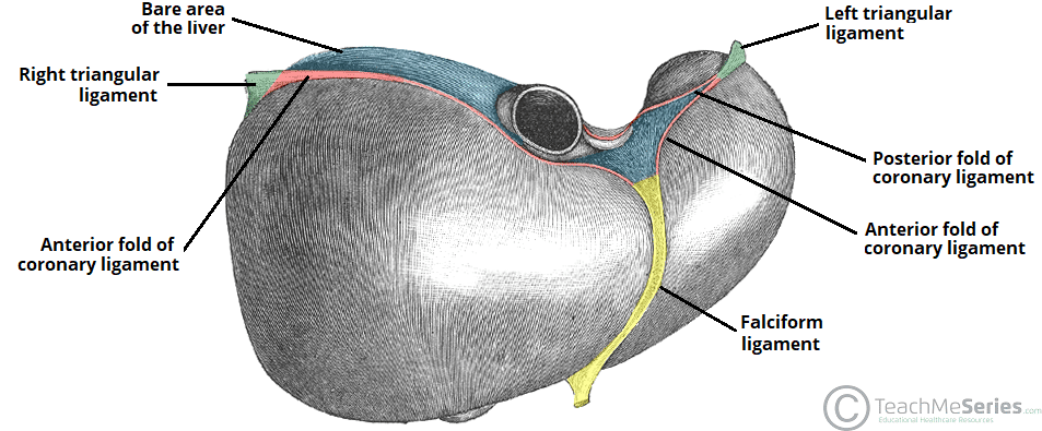
The Liver Lobes Ligaments Vasculature Teachmeanatomy from teachmeanatomy.info In addition researchers at the chair. We cannot see the structure of a virus under a light microscope because it's size is below the resolution capacity of a classical light. Tendons transmit skeletal muscle forces to bone and are essential in all voluntary movement. But at the same time it is interpretive. Tendons and muscles work together to move bones. Using a microscope correctly is a fundamental requirement in any laboratory environment, where sections or microscopic particles have to be examined. Tendons are similar to ligaments; This section is appropriate for students who have no prior experience with using microscopes.
Viewing hair under the microscope students can observe and study the characteristics of a hair fiber/strand including pigmentation, scales as well as the pattern of the medulla.
We cannot see the structure of a virus under a light microscope because it's size is below the resolution capacity of a classical light. Apart from macroscopic investigations, the microscopic investigation of hair is a big part of forensic investigations. Both are made of collagen. Tendons play an important role in the movement by transmitting the contraction force produced by the muscles to the bone they hold, and their contribution to stability to the joints is extremely important. In their relaxed state, the collagen fibers of both tendons and ligaments form a typical wavy pattern, also referred to as a 'crimp,' when viewed under a polarized light microscope. In addition researchers at the chair. Tendons generally have a very complex structure; Cells within the tendons were isolated for analysis. But at the same time it is interpretive. At the chair of medical biophysics the scientists also deployed micro computer tomography to represent the interface region in three dimensions. Transmission electron microscopes an overview. However, tendon cell activity during growth and homeostatic maintenance is less well defined. They are actually heavily composed of connective.
But at the same time it is interpretive. Transmission electron microscopes an overview. This video takes you through microscope images of cells going through mitosis and identifies the different phases under the microscope and on a micrograph. Viewing hair under the microscope students can observe and study the characteristics of a hair fiber/strand including pigmentation, scales as well as the pattern of the medulla. The diagram is very clear, and labeled;
100 Histology Muscle Ideas Muscle Skeletal Muscle Histology Slides from i.pinimg.com Microscope information, images from beneath the microscope and educational science projects. But at the same time it is interpretive. Figure 1.2 light microscope and its parts. However, tendon cell activity during growth and homeostatic maintenance is less well defined. Some of the fibres have been teased apart. Tendons generally have a very complex structure; The diagram is very clear, and labeled; This video takes you through microscope images of cells going through mitosis and identifies the different phases under the microscope and on a micrograph.
This is also a good review for those who could use a refresher on the anatomy of a microscope.
It projects an enlarged and illuminated image o the object to. In their relaxed state, the collagen fibers of both tendons and ligaments form a typical wavy pattern, also referred to as a 'crimp,' when viewed under a polarized light microscope. Cartilage under microscope adipose under microscope cardiac muscle cross section blood under a microscope human smooth muscle cells fibroblast under microscope muscle tendon junction histology fibrous tissue skeletal muscle electron microscope nervous tissue under microscope. Microscope, instrument that produces enlarged images of small objects, allowing the observer an exceedingly close view of minute structures at a scale microscope slides are small rectangles of transparent glass or plastic, on which a specimen can rest so it can be examined under a microscope. Coloured scanning electron micrograph (sem) of tendon fibres. Mnemonics that can be used to remember the anatomy of the ankle tendons from anterior to posterior as they pass posteriorly to the medial malleolus of the tibia under the flexor retinaculum in the tarsal tunnel include: Tenocytes constantly repair small amounts of damage to the matrix under normal circumstances; The enthesis encounters very high mechanical demands and the regenerative capacity is very low resulting in high rupture recurrence rates after. Some of the fibres have been teased apart. Learn vocabulary, terms and more with flashcards, games and other study tools. The substances that can only be seen. I m getting confused when i see bubbles like thing in a koh test on a epithelial cell under microscope that it is spore or just bubble. Tendons generally have a very complex structure;
In addition researchers at the chair. This video takes you through microscope images of cells going through mitosis and identifies the different phases under the microscope and on a micrograph. Microscope, instrument that produces enlarged images of small objects, allowing the observer an exceedingly close view of minute structures at a scale microscope slides are small rectangles of transparent glass or plastic, on which a specimen can rest so it can be examined under a microscope. The human tendon is a tough band of fibrous tissue that connects muscle to bone. Mnemonics that can be used to remember the anatomy of the ankle tendons from anterior to posterior as they pass posteriorly to the medial malleolus of the tibia under the flexor retinaculum in the tarsal tunnel include:
Skeletal Muscle Tissue Histology Kenhub from thumbor.kenhub.com Tendons play an important role in the movement by transmitting the contraction force produced by the muscles to the bone they hold, and their contribution to stability to the joints is extremely important. Figure 1.2 light microscope and its parts. Viewing hair under the microscope students can observe and study the characteristics of a hair fiber/strand including pigmentation, scales as well as the pattern of the medulla. This video takes you through microscope images of cells going through mitosis and identifies the different phases under the microscope and on a micrograph. Under the light microscope, the tendons of patients suffering jumper's knee do not consist of tight parallel collagen bundles but instead are separated by increased mucoid ground substance that gives them a disorganised and discontinuous appearance. Mnemonics that can be used to remember the anatomy of the ankle tendons from anterior to posterior as they pass posteriorly to the medial malleolus of the tibia under the flexor retinaculum in the tarsal tunnel include: The substances that can only be seen. We cannot see the structure of a virus under a light microscope because it's size is below the resolution capacity of a classical light.
Electron microscopy of cultured epidermal ebs 2117 cells reveals.
In addition researchers at the chair. Mnemonics that can be used to remember the anatomy of the ankle tendons from anterior to posterior as they pass posteriorly to the medial malleolus of the tibia under the flexor retinaculum in the tarsal tunnel include: This is also a good review for those who could use a refresher on the anatomy of a microscope. Some of the fibres have been teased apart. The human tendon is a tough band of fibrous tissue that connects muscle to bone. Both are made of collagen. Microscope information, images from beneath the microscope and educational science projects. Tendons are similar to ligaments; The diagram is very clear, and labeled; Microscopes work on the physical principle of magnification where the image of an object is magnified so that it can be visible. Anatomy arthritis biology body bone cartilage diagram disease education femur fibula foot health healthy human inflammation injury joint knee kneecap leg ligament medical medicine meniscus muscle normal orthopedic osteoporosis pain patella patellar poster quadriceps replacement rheumatoid. The microscope is perhaps one of the most fundamentally important pieces of equipment that you will use in the laboratory environment. Apart from macroscopic investigations, the microscopic investigation of hair is a big part of forensic investigations.
Microscopes work on the physical principle of magnification where the image of an object is magnified so that it can be visible tendon diagram. Tenocytes constantly repair small amounts of damage to the matrix under normal circumstances;

Comments
Post a Comment